

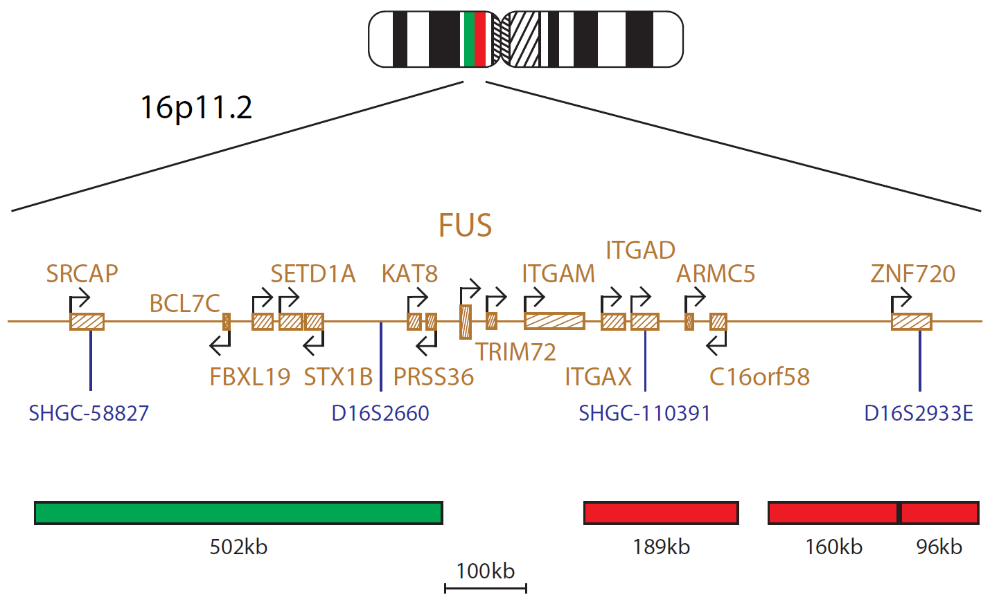
The FUS Breakapart probe consists of one 502kb probe labelled in green, situated distal to the FUS gene and covering markers SHGC-58827 and D16S2660, and three probes (189kb, 160kb and 96kb) labelled in red, situated proximal to the FUS gene and covering markers SHGC-110391 and D16S2933E.
The FUS (FUS RNA binding protein) gene at 16p11.2 is a member of the FET family of protein-encoding genes, closely-related to the EWSR1 (EWS RNA binding protein 1) gene1.
Recurrent rearrangements involving the FUS gene with a number of different partner genes have been reported in various types of neoplastic disease, notably soft tissue sarcomas and acute myeloid leukaemia. In some tumour types FUS and EWSR1 may replace each other as fusion partners2.
In soft tissue sarcoma, approximately 90% of cases of myxoid liposarcoma are characterised by the presence of a FUS-DDIT3 rearrangement arising from a t(12;16)(q13;p11) translocation3,4; the FUS-CREB3L1 and the FUS-CREB3L2 fusions, resulting from t(11;16) (p11;p11) and t(7;16)( q32-34;p11) translocations respectively are characteristic of low-grade fibromyxoid sarcoma5, whereas the t(12;16)(q13;p11) translocation resulting in a FUS-ATF1 fusion gene is seen in angiomatoid fibrous histiocytoma6.
This breakapart probe has been designed to allow detection of FUS rearrangements regardless of the partner gene involved.
The quality and reproducibility of results using the CytoCell kit has been vital in accurately detecting co-deletions in our glioma investigations. We now have a cost-effective test that we can rely on that is also easy to use and interpret. We've been consistently impressed with this kit - not to mention the support offered by OGT's customer service, and have completely transitioned over to CytoCell probes.
Gavin Cuthbert, FRCPath
Head of Cancer Cytogenetics, Northern Genetics Service, Newcastle, UK

 Visit USA site
 Visit Canada site


